Pseudocalcium clustering of retinal ganglion cells
In this work we aimed to cluster the extracellular activity of retinal ganglion cells(RGCs) based on the cluster definition of the landmark study carried out by Baden et al in 2016 [1]. Since there are still some technical limitations in imaging the electrical activity of neurons during electro-stimulation with calcium imaging techniques, we sought to record light and electrical induced activity of RGCs with multi-electrode array(MEA) and classify them using feature set provided by Baden et al. These results can help us to better characterize the electrical properties of RGCs under electro-stimulation for bionic vision research.
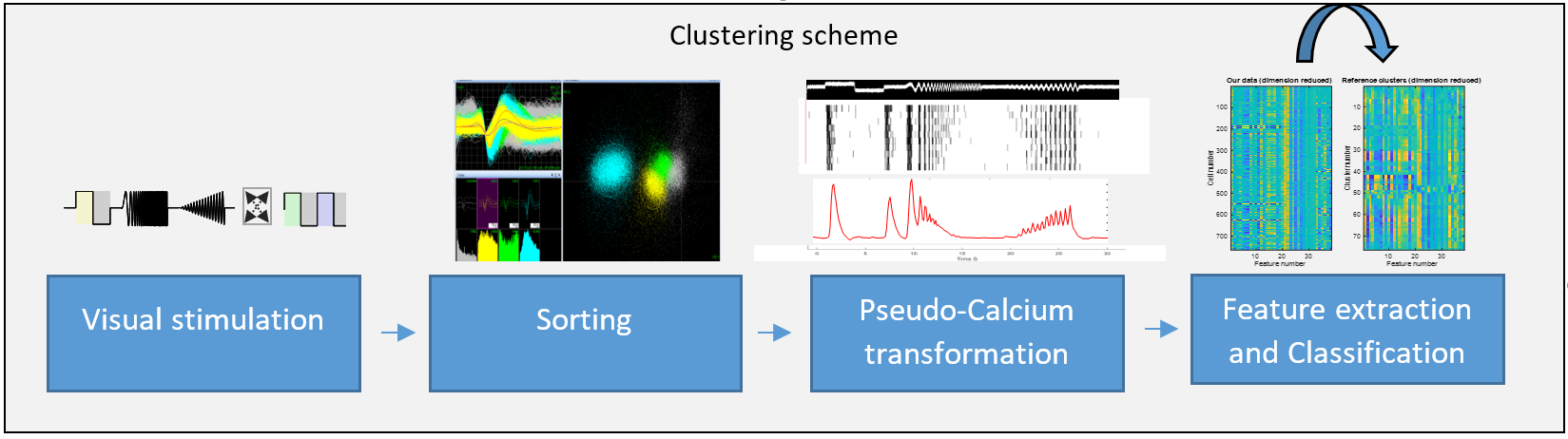
Figure 1. The proposed clustering workflow.
Click here to see the high quality image
In order to match the two clustering methods, the spiking data were first converted into pseudocalcium signals using the OGB-1 convolution kernel (figure 1). After converting data to pseudocalcium signals, we projected them into lower dimensions using the sparse principle component features of the reference data. This allowed us to analyze our data according to the cluster definition of the Baden study. After computing the distance matrix each cell was assigned to the most similar cluster (figure 2).
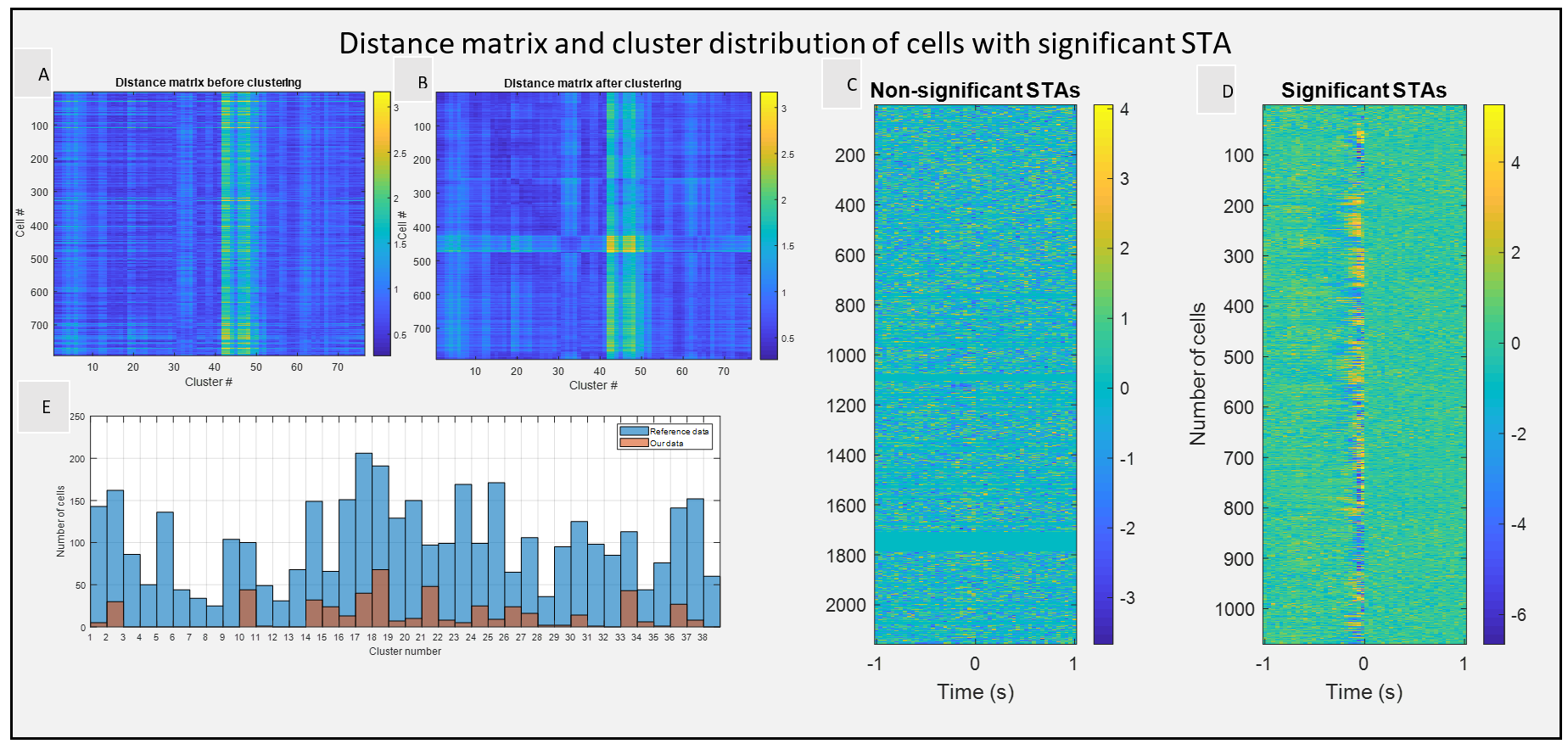
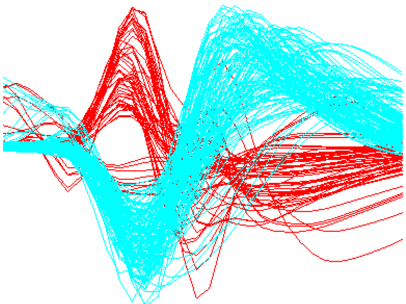
Figure 2. (Left) Distance matrix of pseudocalcium data before and after clustering. (Right) Electrical input filters with significant deflection.
Click here to see the high quality image
Although most of our data were matched to the cluster groups of the reference study by using pseudocalcium signals, some groups failed to match, specifically the clusters with sustained chirp response.
This might be due to the subthreshold calcium signals that are not led to spike generation during low contrast or high-frequency stimulation periods.
Therefore, there is the possibility that some of the cells that belonged to sustain clusters were misclassified as transient groups (figure 3).
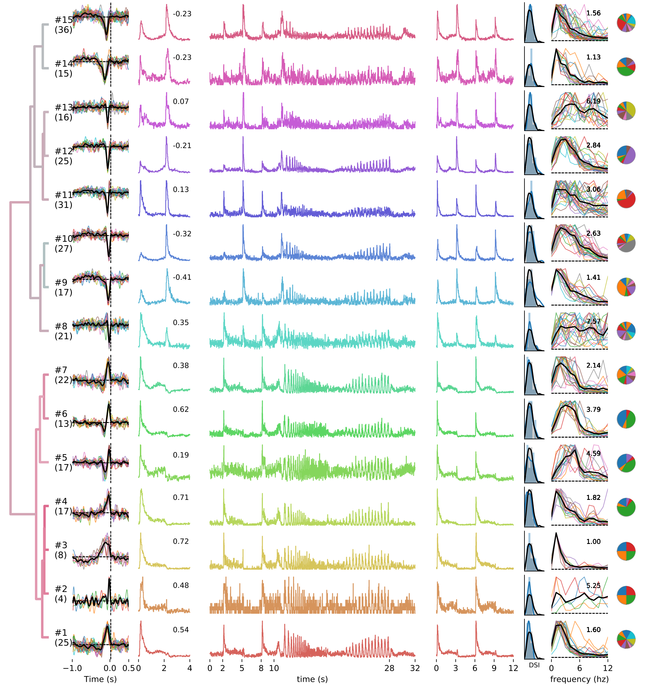
Figure 3. Clustered RGCs. (Left) reference data, (middle) our data with 653 neurons, (right) our data 2303 neurons.
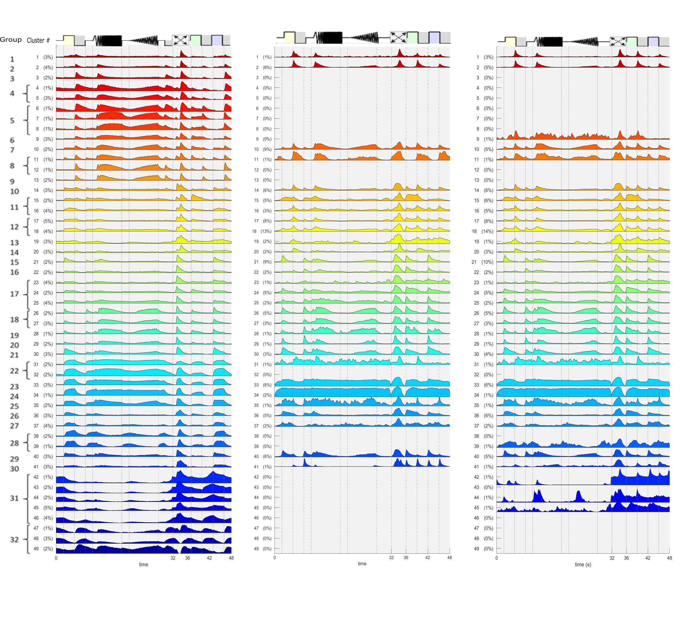
Click here to see the high quality image
By assigning the light-evoked activity of each RGC to one of the defined groups, our data was found to match 27 of the 32 defined groups.
Ultimately, we concluded that due to its shortcomings, the pseudocalcium clustering framework might not be the best solution and other alternatives may have a better performance. In another effort, we implemented a clustering framework based on spike train distance metrics to characterize the electrical properties of RGCs under electro-stimulation. The results of that work can be found here .
Supervisors:: Dr. Daniel Rathbun, Prof. Eberhard Zrenner.