Clustering visual and electrical response of RGCs using spike distance metrics.
Current retina implants implement pulsate stimuli to activate the neural circuits of the retina. this type of stimulation can activate antagonist retinal pathways which lead to the improper perception of the visual scenes. Developing a precise stimulation strategy with the ability to preferentially target the retinal neural circuit is one of the alternative methods to improve the accuracy of restored vision.
For this project, we recorded extracellular activity from RGCs of more than 30 mice using multi-electrode arrays. We used the electrical Gaussian noise stimulation to characterize the response properties of different ganglion cell types in the retina. In addition, a variety of visual stimuli including moving bars, contrast and temporal frequency chirps, blue-green color, and flashes were used to classify the recorded ganglion cells based on their light-evoked activity and assess their electrical properties (see figure 1).
Figure 1.experimental setup. MEA recording setup (top), visual and electrical stimulus sets(bottom).
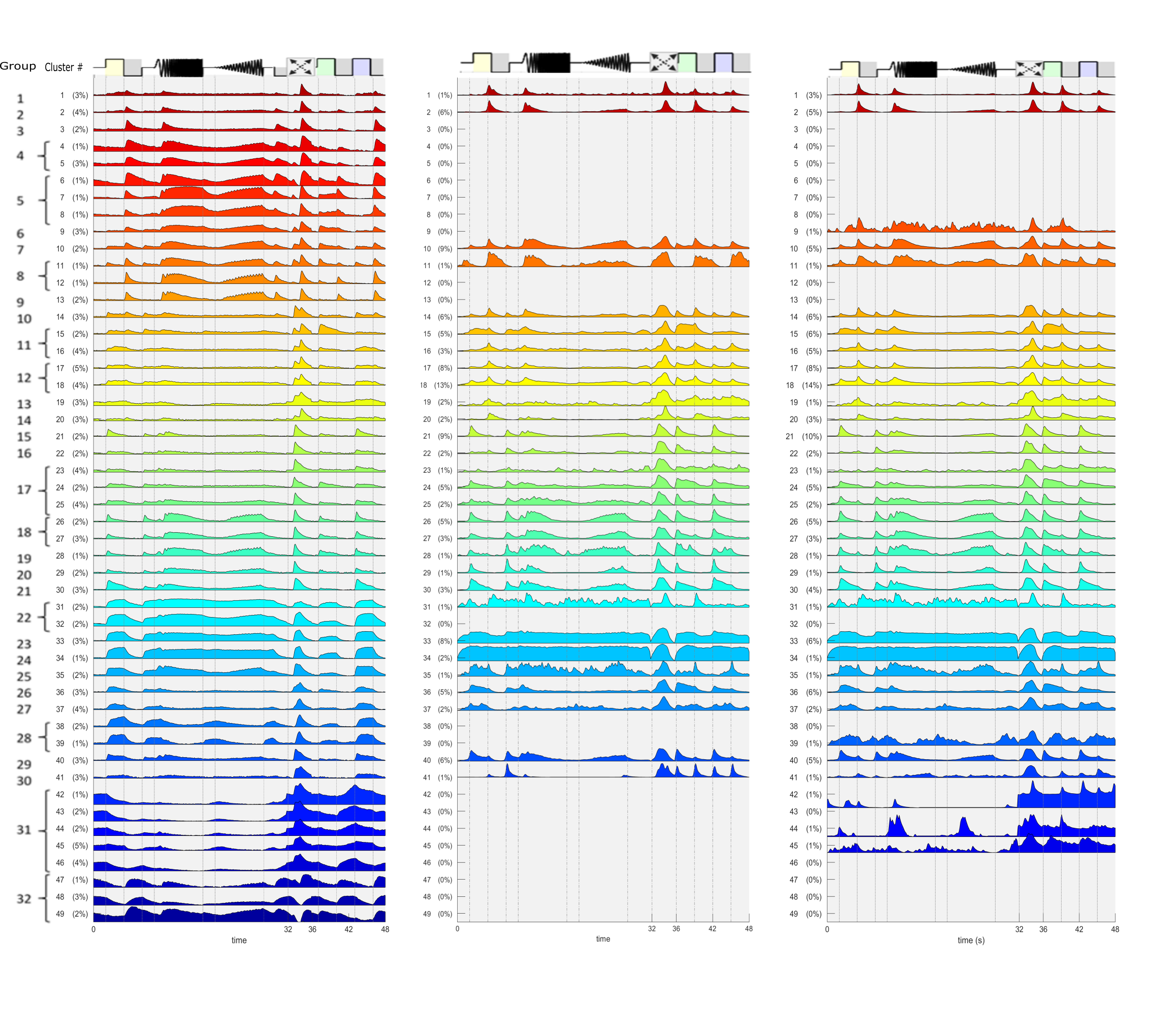
We used a non-parametric clustering framework to cluster visual and electrical induced activity of RGCs [1]. in the first step, the spike timed based distance matrix of cells was calculated [2] and clustered via a hierarchical clustering algorithm. The clustering algorithm detected 37 clusters, including ON, OFF, and ON-OFF cell types (figure 2). By calculating the average temporal electrical receptive field of each cluster we observed a notable correspondence between the cell types and their electrical signature.
Figure 2. Clustering RGCs based on light-induced activity. (a) cluster dendrogram. (b-d) light-evoked activity (e) disribution of direction selectivity index (f) Electrical input filter.
By taking the advantage of reverse correlation analysis coupled with machine learning approaches, we were able to establish a full catalog of electrical profiles for different ganglion cell types. We showed that ON and OFF cell types reflect dissimilar electrical transfer functions generated by different retinal pathways.
In the second step, we clustered cells based on the shape of their electrical profiles. We detected both upward and downward liner filters with a wide range of variety in their widths and latencies, which can represent the electrical characteristics of different cell types (see figure 3). We observed that clusters with upward linear filters contain only the activity of ON cells, whereas clusters whose linear filters showed a downward deflection contained only the activity of OFF and ON-OFF cells. This notable difference between the light-induced activity of clusters with upward and downward electrical profiles shows that subthreshold activation of the retina may activate separate retinal pathways.
Figure 3. Clustering RGCs based on the shape of electrical input filters. (a) cluster dendrogram. (b) Electrical input filter, (c-e) light-evoked activity (f) disribution of direction selectivity index (g) Power spectrum density.
Moreover, pharmacological manipulation of degenerated retina revealed that input filters with downward deflection were disrupted by blocking the synaptic connections between photoreceptors and On-bipolar cells, while the electrical input filters with upward deflection were subjected to smaller changes after blocking this circuit (not shown).
The results of this study deepened our insight into the underlying mechanism of subthreshold electrical activation of retinal pathways. First of all, we showed RGCs can be stimulated via integrated subthreshold stimulation. In addition to that, we showed that ON and OFF pathways have different electrical input filters which may provide the possibility of selective activation.
Supervisors: Dr. Daniel Rathbun, Dr. Zohreh Hosseinzadeh, Prof. Eberhard Zrenner.